

antscan is a pilot project for creating digital libraries of 3D invertebrate anatomy with high-throughput synchrotron micro-CT imaging
The Project

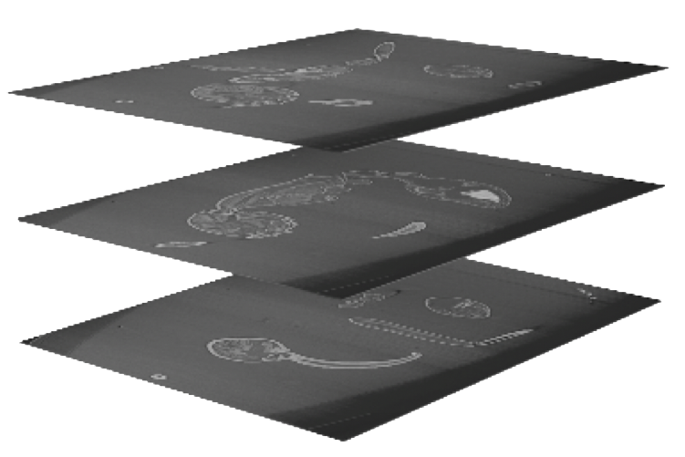
Methods and Workflow
Learn more about the specifics of Serial Synchrotron CT-Scanning and how we organized thousands of ants for imaging
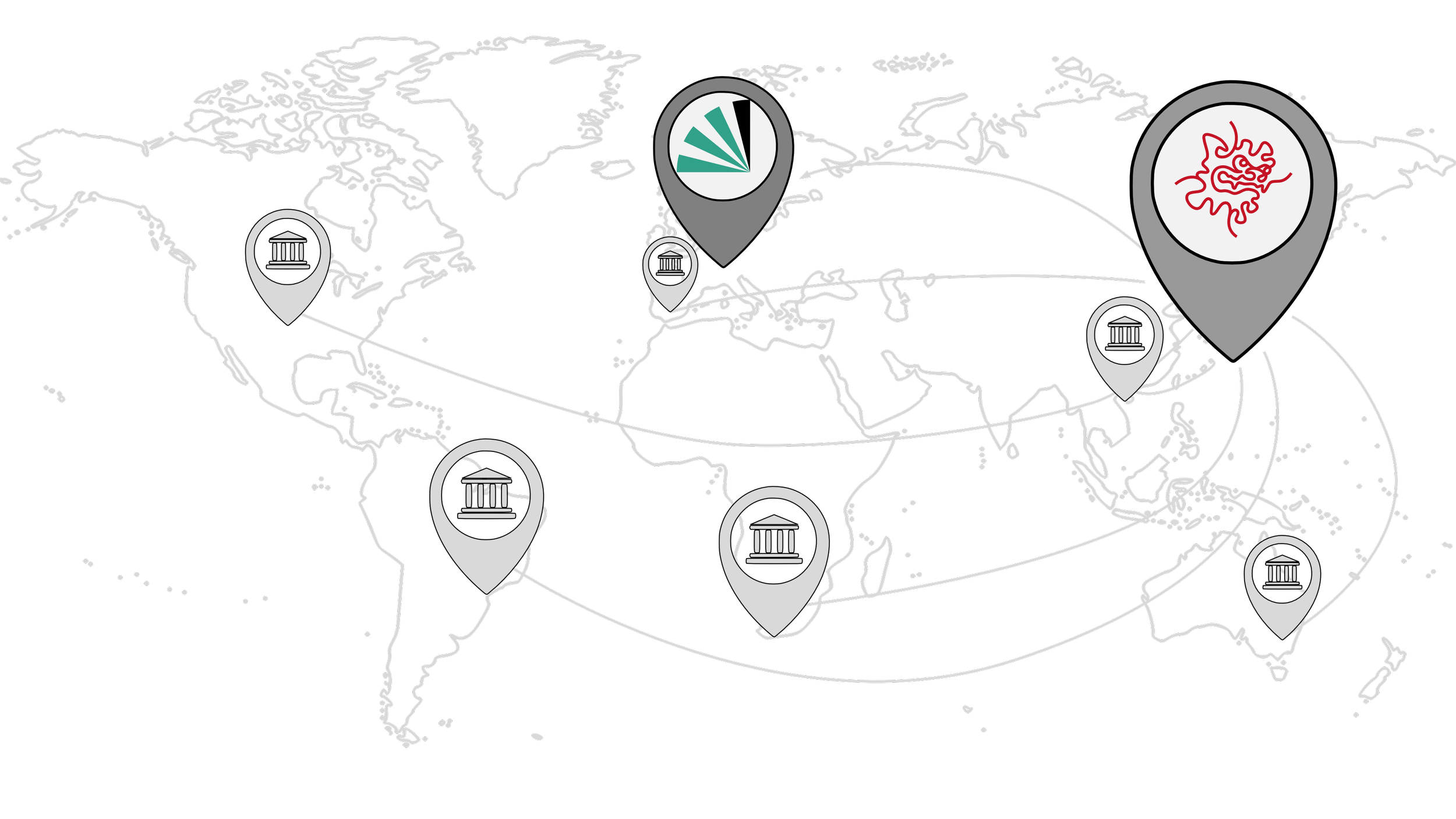
In antscan, we use serial synchrotron X-ray microtomography to capture 3D ant morphology with high throughput. Ants are diverse, ecologically dominant, and functionally important. Through studying anatomy, we investigate bridges between genomes and environment, and we can learn more about adaptations and the effects of natural selection. The result of the project is an open-source dataset, currently consisting of ~2200 scanned specimens from ~900 species and 210 genera with each scan acquired in a minute. The set of species covers the ant tree of life, has a global geographic scope. In parts, the 3D morphological data is directly paired with ongoing ant genome sequencing efforts. The antscan project is a first large-scale, comprehensive library of skeletal and soft-tissue for a diverse clade. We hope antscan will be part of a new era of high-throughput imaging-based studies on the evolution, structure, and function of organismal phenotypes.
Scans
Explore the list of ants and find the link to
the Biomedisa antscan database

Antscan is supported by
