Discover below the building blocks of antscan described in plain language. For more details, dive deeper into the scientific paper presenting the antscan project.

To make antscan happen, at OIST, we organized thousands of ant specimens from around the world to get a good sampling of global biodiversity. This can only happen through global coordination and collaboration. At the KIT Light Source, the Serial X-ray Microtomography technique was developed and so far, it is the only Synchrotron facility that allows such a project. Finally, on Biomedisa supported by the KIT and University of Heidelberg University data centers we can freely offer terabytes of ant scans for download. (Thomas van de Kamp, Julian Katzke)
The ants in antscan
Where do they come from?
In antscan, we have the ambition to adequately digitize the morphological side of ant biodiversity. Because there are more than 15,000 described species and ants are globally distributed, the antscan project needs to reflect that diversity. Therefore, museum curators and ant scientists from around the world come together in antscan to provide preserved ant specimens for 3D imaging.
How to preserve ant anatomy?
The two main ways in science to store and preserve insects are either pinning them or storing them in alcohol. Gluing ants on a fine piece of paper and putting the paper on a metal makes them very easy to observe under a microscope and therefore, they are invaluable for measuring ants and describing new species. This works because the ants’ exoskeleton can stay intact even when the ant is all dried up. With antscan 3D data however, we also want to be able to look at what’s on the inside. Conveniently, simply storing insects in alcohol preserved many soft tissues, even over many years when treated properly.
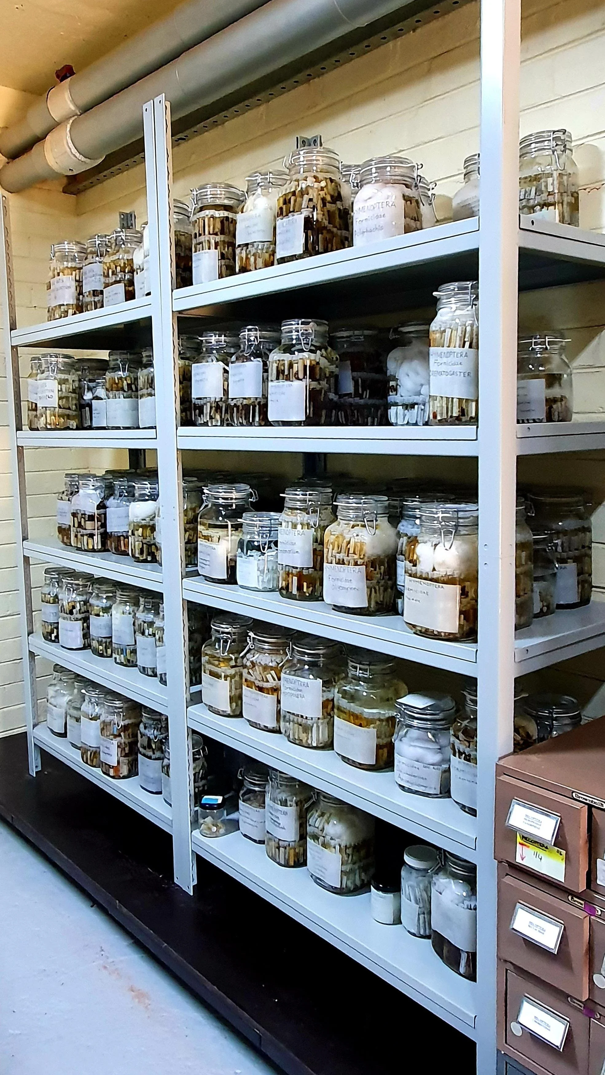
Ethanol and other spirit-preserved specimens at the Australian National Insect Collection in Canberra. (Francisco Hita Garcia)
3D-imaging thousands of ants
CT
Computed Tomography (CT) is a technique to create 3D images from sampling 2D projections of a 3D volume. If we want to digitize internal details, we can use Xray images as the projections. In medical CT in hospitals, the source of Xrays spins around the patient to get a CT scan. In μCT, the object itself is rotated while source and camera are stationary.
Synchrotron
At the Synchrotron Light Source in the Karlsruhe Institute of Technology (KIT), a storage ring produces radiation for various applications by accelerating electrons. This Synchrotron radiation is used at the KIT for CT among other things. At so called beamlines, the Synchrotron radiation is harvested. The KIT μCT beamlines are optimized to translate the high intensity of the Synchrotron Xrays into extremely low exposure times when sampling projections.
This animation shows the Xray projections from which the 3D volume is reconstructed. The high intensity of the synchrotron allows exposure times of 0.01 seconds. The camera operates at 70-100 frames per second and in total, 3000 images are taken per specimen. (KIT, Julian Katzke)
Serial Synchrotron μCT
To achieve the high-throughput necessary to tackle thousands of CT scans at a time, at the KIT, the potential for fast Xray sampling is met with more technological innovation. Rapid μCT scanning of many samples is achieved with a robotic sample changing setup, high speed cameras to document the Xray images, and fast network connections to efficiently transfer the data. Separate from raw data acquisition, the samples are then processed into 3D tomograms.